Category: Parkinsons Disease Information(Page 1 of 16)
Access 20 years of research focused on identifying natural approaches, therapies and treatments that offer the opportunity to celebrate relief from the symptoms associated with a diagnosis of Parkinsons disease.
This brief video explains the purpose and goal of the Parkinsons Recovery Six Month Recovery Program
Two opportunities to enroll in the Six Month Recovery Program: a monthly subscription and a one time payment. Visit the links below for more information about each:
1. Initial strategy session with me personally to flesh out the potential causes of your symptoms to identify therapies and treatments that will successfully address them.
This is not some surface-level consultation. It is a deep dive into your specific history and situation. We evaluate what you have tried that did not succeed and what did help. We consider new options.
2. Together we lay out a custom-crafted program of recovery created specifically for your situation.
3. We connect with hour long depth sessions twice a month for six (6) months with a video type interface similar to Zoom or with phone calls if that is more convenient.
4. Together we review all the options given the probable cause of your symptoms and focus on therapies that offer the greatest possibility of success. The program of recovery is re-evaluated and adjusted as needed from session to session.
During our sessions every two weeks we …
Review your progress since our last session together
Identify and solve any roadblocks you confronted
Refine strategies based on real-world results documented by research
Help track and celebrate your progress
Accelerate your progress with options documented by research proven to offer symptom relief
5. Unlimited email access to me personally so we can always be in touch between sessions.
6. Enrollment in the Parkinsons Recovery® Jump Start to Wellness course is included.
Guarantee
The goal of these bi-weekly sessions for six months is for you to celebrate a relief from your symptoms. If for any reason you do not experience the relief from symptoms you anticipated after six months, 80% of all subscription payments will be refunded.
Note: As a researcher, not a medical doctor, I do not address issues with prescription medicines. Your medical doctor is the go to person to sort these issues out. We focus on natural options that offer the opportunity to recover.
Robert Rodgers PhD Founder 2004 Parkinsons Recovery® Road to Recovery from Parkinsons Disease
I offer two insights on how to manifest recovery. These are not guesses. My recommendations are inspired by interviews with hundreds of individuals who report on what they do to to celebrate a successful journey on the road to recovery.
First. A surprising number of factors contribute symptoms. Focusing on one and only one takes you a little way down the road to recovery, but not far! Individuals who celebrate success embrace a combination of therapies that address the factors that are causing their symptoms. The average is 5-6 different approaches.
Inviting an eclectic group of persons to be members of your medical team is a smart move. By this I mean medical professionals who offer different approaches to recovery. Expand your approach behind physical remedies. Include those that clear dark emotions, remove toxins and release the grip of past traumas (physical as well as emotional).
Approach the challenge of recovery by accepting the healing potential offered by different modalities. This transforms despair and hopelessness to rejuvenation and regeneration.
Second. Persistence pays off in the long run. Many people begin a tried and true natural therapy backed up by extensive research and begin feeling worse or they are simply not feeling better. They conclude the therapy is not working (and maybe even making them worse off) so they abandon it for good reason. This therapy was supposed to help you feel better and you experienced the undesirable outcome of feeling worse or the unwelcome outcome of no improvement. Really?
Really. When the body is healing from the inside out you will feel often worse in the beginning. The road to a successful recovery for everyone is rocky – full of starts, stops and even what seems to be stagnation. It took years for neurological symptoms to surface. Why expect they will resolve in a few months?
A strategy of persistence will pay off in the long run.
I recommend you first do a scan of the natural therapies that are supported by sound research evidence. There are a surprising number of ground breaking therapies that have surfaced over the last few years that use light and sound to heal the body from the inside out. Check in with your wisdom and intuition to decide which ones would be the best for you to embrace.
When you are drawn to pursue one of these therapies it is because your body is telling you it will help you heal. Acknowledge the long term costs involved. The positive outcome you anticipate may only manifest a year or longer after starting the therapy. Once you decide on a therapy, go for it.
Persist. Yes, you will likely feel worse one day or another. Some days will likely be much better than others. When you do feel worse, say to your self – great. I know my body is finally healing from the inside out.
There are of course options that help symptoms reverse temporarily like medicines. That will not be the case with most of the ground breaking therapies that support the body’s ability to return to its natural state – one of balance and harmony. The body does know how to heal. Just give it the support it needs and hang in there.
My second recommendation is take your time deciding which natural therapies to pursue. When you make a decision to pursue a natural therapy – persist. Focus and commitment are the keys to a successful recovery program.
Researchers have found conclusive evidence that symptoms associated with a diagnosis of Parkinsons originate in the gut. Many persons in my audience report having digestive problems such as constipation. But what causes digestive problems? Mouth health affects gut health.
The vagus nerve is a highway that connects the gut with the brain. Infections in the gut have readily accessible access to the brain. Sores and infections in the mouth travel to the gut. These infections in the gut travel to the brain. No one can dispute the connection.
Let me ask you three questions …
Do you often get mouth sores?
Having difficulty clearing a gum infection?
Is your digestion compromised?
If the answer is yes to any of these questions, I recommend you begin using a natural mouth wash that contains no synthetic chemicals, nothing harsh. What you want is a cleanse that contains ingredients your body recognizes, not a mouthwash with harmful chemicals that can not even be swallowed.
The mouth cleanse that meets these conditions is Suvia. Suvia isn’t just another mouthwash that can never be swallowed. It’s a hydrogel-based oral rinse made from 100% natural food ingredients, designed specifically for soft tissue health, not just surface cleaning.
Summary. Resolve infections and sores with a natural mouth cleanse to celebrate an improvement in mouth health and, in turn, your digestion. When digestion improves, studies find that neurological symptoms will accompany the improvement.
As my video preview below explains, studies report that Cannabidiol CBD offers relief from Parkinson’s symptoms. Annual Parkinsons Recovery surveys also report success with reversing Parkinson’s symptoms when taking CBD oil. It is one of the most frequently listed therapies reported to be useful to members of the Parkinsons Recovery audience.
CBD stands for “cannabidiol oil”. It is extracted from either the Hemp plant or the Marijuana plant. Using CBD allows the medical benefits of medical marijuana without the high that is associated with THC. CBD in itself has no psychedelic properties and is considered safe for consumption
Abstracts of recent CBD studies and posted at the bottom of this post. Taken together, studies find that CBD …
Protects dopaminergic neurons
Reduces neuro-inflammation and oxidative stress
Improves motor symptoms
Increases REM sleep
Reduces insomnia episodes
Enhances mood
Reduces fatigue
Reduces chronic pain
Helpful for stroke victims.
If taking the Alovea Entourage CBD does not provide the relief from symptoms anticipated, contact Robert for a full 100% refund. Offer available to USA residents only. robert@parkinsonsrecovery.com
About Alovea Entourage Cannabidiol CBD
Alovea Entourage contain plant-derived cannabinoids, distinct from endocannabinoids that are produced in the body and synthetic cannabinoids. It includes Acemannan, an extract from the Aloe plant, to help the body’s management of mood, memory, sleep and joint health.
SYMBIOTIC IMMUNE SUPPORT
There are many products that have been formulated with additional ingredients to provide support for specific benefits like improved sleep, mood and joint health. One of the most profound impacts has come from the addition of immune modulating aloe vera Acemannan.
Combining the immune modulating fractions of Acemannan with high quality CBD hemp oil provides a symbiotic impact (1 + 1 = 4) that can dramatically enhance overall immune support.
PATENT-PENDING FORMULA INCLUDES:
Organic Broad Spectrum Hemp Oil
Super critical CO2 extraction
Each batch tested to ensure 0.0% THC
Absorption increased by 5-7 times over other hemp oil products with water compatible nanotechnology
Enhanced Entourage Effect through proprietary terpene blend
Symbiotic effect with the highest rated immune modulating Alovéa AE (Acemannan Extract)
Wonderful cinnamon and tropical flavors!
Recent CBD (Cannabidiol) Research Studies
Clinical research on CBD (cannabidiol) includes studies of anxiety, cognition, movement disorders, and pain. Abstracts of recent studies are included below.
Pharmaceuticals (Basel). 2026 Feb 17;19(2):330. Cannabidiol in Neurology: Current Insights and Translational Perspectives
Abstract
Cannabidiol (CBD) is one of the most studied compounds of Cannabis sativa and has attracted significant interest due to its therapeutic and beneficial properties, which have been confirmed in numerous preclinical and clinical studies over the last few years.
A great advantage of CBD over the other widely known Cannabis sativa ingredient, ?-9-tetrahydrocannabinol (THC), is that CBD does not exert intoxicating and psychoactive effects, making it an attractive candidate for therapeutic applications in neurological disorders.
CBD has been shown to exert antioxidant, analgesic, anti-inflammatory, and neuroprotective effects, with therapeutic potential for various neurological conditions. To date, the only drug that consists solely of highly purified CBD is Epidiolex, which is used in the management of severe forms of epilepsy such as Dravet syndrome and Lennox-Gastaut syndrome. Another legal medication containing CBD (albeit with the addition of THC) is Sativex, used to alleviate spasticity in multiple sclerosis.
Besides epilepsy, preclinical data suggest that CBD alone may be potentially beneficial in treating chronic pain, multiple sclerosis, Alzheimer’s and Parkinson’s diseases, or stroke.
The safety profile of CBD is generally considered favorable, as the most commonly reported adverse effects are mild (e.g., somnolence, diarrhea).
******
Cannabis Cannabinoid Res. 2026 Feb;11(1):11-29. Unveiling Neurological Benefits: A Review of Hemp Leaf, Flower, Seed Oil Extract, and Their Phytochemical Properties in Neurological Disorders
Abstract
Neurological disorders such as epilepsy, Alzheimer’s disease, Parkinson’s disease, and multiple sclerosis present significant global health care challenges, with complex pathophysiology and limited therapeutic options that often carry substantial side effects.
Hemp-derived compounds, particularly from Cannabis sativa seeds, leaves, and flowers, have gained attention for their potential neuroprotective properties. This review aims to synthesize the current evidence surrounding the therapeutic benefits of hemp-derived compounds, focusing on their bioactive phytochemical profiles, mechanisms of action, and therapeutic efficacy in treating neurological disorders.
A comprehensive review of pre-clinical and clinical studies was conducted, analyzing the phytochemical composition of hemp extracts, including cannabinoids (such as cannabidiol, CBD), terpenes, flavonoids, and polyunsaturated fatty acids. We explored their mechanisms of action through interactions with the endocannabinoid system, neurotransmitter receptors, inflammatory pathways, and oxidative stress mechanisms.
The review highlights the therapeutic potential of hemp-derived extracts in mitigating various neurological conditions. Pre-clinical and clinical studies have demonstrated their efficacy in reducing seizure frequency in epilepsy, protecting dopaminergic neurons in Parkinson’s disease, alleviating neuroinflammation and oxidative stress in Alzheimer’s disease, and promoting remyelination in multiple sclerosis.
The entourage effect, where cannabinoids, terpenes, and flavonoids work synergistically, enhances these therapeutic effects. Innovations in extraction technologies have optimized yield and preserved bioactivity, further enhancing clinical relevance.
Hemp-derived compounds exhibit significant neuroprotective and therapeutic potential for managing neurological disorders.
******
J Clin Sleep Med. 2025 Jan 1;21(1):69-80. Effects of a cannabidiol/terpene formulation on sleep in individuals with insomnia: a double-blind, placebo-controlled, randomized, crossover study
Abstract
Study objectives: Cannabidiol (CBD) is increasingly used as a health supplement, though few clinical studies have demonstrated benefits. The primary objective of this study was to evaluate the effects of an oral CBD-terpene formulation on sleep physiology in individuals with insomnia.
Methods: In this double-blind, placebo-controlled, randomized clinical trial, 125 individuals with insomnia received an oral administration of CBD (300 mg) and terpenes (1 mg each of linalool, myrcene, phytol, limonene, ?-terpinene, ?-terpineol, ?-pinene, and ?-caryophyllene) for ? 4 days/wk over 4 weeks using a crossover design. The study medication was devoid of ?9-tetrahydrocannabinol. The primary outcome measure was the percentage of time participants spent in the combination of slow-wave sleep (SWS) and rapid eye movement (REM) sleep stages, as measured by a wrist-worn sleep-tracking device.
Results: This CBD-terpene regimen marginally increased the mean nightly percentage of time participants spent in SWS + REM sleep compared to the placebo (mean [standard error], 1.3% [0.60%]; 95% confidence interval, 0.1-2.5%; P = .03).
More robust increases were observed in participants with low baseline SWS + REM sleep, as well as in day sleepers. For select participants, the increase in SWS + REM sleep averaged as much as 48 minutes/night over a 4-week treatment period. This treatment had no effect on total sleep time, resting heart rate, or heart rate variability, and no adverse events were reported.
Conclusions: Select CBD-terpene ratios may increase SWS + REM sleep in some individuals with insomnia and may have the potential to provide a safe and efficacious alternative to over-the-counter sleep aids and commonly prescribed sleep medications.
******
J Obstet Gynaecol Can. 2025 Jul;47(7):102839. Cannabidiol Use and Perceptions of Effectiveness in Women With Chronic Pelvic Pain
Abstract
Objectives: To investigate cannabidiol (CBD) use in women with chronic pelvic pain and nociplastic pain, as represented by a diagnosis of fibromyalgia, including impact on pain and associated symptoms, substitutions for other medications, and side effects.
Methods: This was a cross-sectional study using data derived from an online survey distributed through the National Fibromyalgia Association between April and May of 2020. This secondary analysis was limited to participants with concurrent chronic pelvic pain diagnoses. Participants were asked about their history of CBD use and sub-grouped by use patterns.
Results: Of 1382 included participants, 501 (36.3%) were in the “Never Use” group, 404 (29.2%) were in the “Past Use” but not “Current Use” group and 477 (34.5%) were in the “Current Use” group. Of 477 participants in the “Current Use” group, 80.9% (n = 386) reported pain improvement. The most common indications for current CBD use were pain, insomnia, and anxiety. Participants who reported pain improvement were more likely to report improved sleep (P < 0.001), anxiety (P < 0.001), depression (P = 0.001), fatigue (P < 0.001), overall health (P < 0.001), and substitution of CBD for at least 1 other medication (P = 0.003) than those who reported no pain improvement. About half of the participants reported side effects, most of which were minor with the most common being drowsiness (n = 138, 29%).
Conclusions: Two-thirds of participants with concurrent chronic pelvic pain and fibromyalgia have used CBD. Improved pain with CBD use correlated with improvement in other symptom domains, such as sleep, mood, fatigue, and overall health. A large proportion of those using CBD reported substituting it for higher-risk medications.
Cannabis Cannabinoid Res. 2023 Oct;8(5):716-730.Cannabinoids in Treating Parkinson’s Disease Symptoms: A Systematic Review of Clinical Studies
Abstract
Background: Parkinson’s disease (PD) is a serious neurodegenerative condition impacting many individuals worldwide. There is a need for new non-invasive treatments of PD. Cannabinoids in the form of cannabidiol (CBD) and delta-9-tetrahydrocannabinol (THC) may offer utility as treatment, and our objective was hence to conduct a systematic review regarding the clinical evidence for the efficacy and safety of cannabinoids in treating PD.
Methods: Screening, data extraction, and quality assessments were all conducted by multiple reviewers, with discrepancies resolved by consensus.
Results: After conducting searches in 4 different databases, 673 articles were screened. Thirteen articles were deemed eligible for inclusion in this review. It was shown that cannabis, CBD, and nabilone (a synthetic form of THC) were capable of consistently improving motor symptoms more than a placebo. All treatments improved various non-motor symptoms, particularly with cannabis improving pain intensity, and CBD improving psychiatric symptoms in a dose-dependent manner. Adverse effects were usually minor, and, in the case of CBD, rare (except at very high doses).
Conclusion:Cannabinoids have been shown to safely offer important potential in treating motor symptoms in PD and some non-motor symptoms.
As a ground breaking discovery, far Infrared treatments offer hope for individuals confronting chronic conditions who have not succeeded in finding ways to reverse their symptoms. Below is a 20 minute interview with CEO of Gladiator Therapuetics Bill Grovatt who offers a fascinating summary of the history behind using far infrared technology in new and innovative ways to treat a variety of chronic conditions.
Gladiator’s far infrared Technology holds potential for chronic conditions to:
Improve mitochondrial function
Reduce neuroinflammation
Enhance overall cellular health and function, which offers the opportunity to mitigate motor and non-motor symptoms
Testimonial from Harvey Caine Please wait – it takes a few minutes for the video to load!
Research on Far Infrared treatments for Parkinsons
Curr Issues Mol Biol. 2023 Dec 6;45(12):9823-9837. Amelioration of Motor Performance and Nigrostriatal Dopamine Cell Volume Using a Novel Far-Infrared Ceramic Blanket in an A53T Alpha-Synuclein Transgenic Parkinson’s Disease Mouse Model
Abstract
We had attended a Parkinson’s Disease (PD) patient for a non-healing wound who reported a marked decrease in his hand tremor and freezing of gait when his wound was exposed to a ceramic far-field infrared (cFIR) blanket.
Efforts to slow or stop symptoms have resulted in the development and use of dopaminergic drug replacement therapy. Unfortunately, there is a loss of about 70-80% of substantia nigral dopaminergic neurons in patients by the time they are diagnosed with PD, and various dopaminergic drugs provide only temporary relief of their motor symptoms. There are limitations in treating PD with many conventional medications, necessitating a combination of pharmaceutical and non-pharmacological therapy as an essential adjunct to better address the health and welfare of PD patients.
We used male adult A53T alpha-synuclein transgenic mice exposed to a ceramic far-infrared blanket. Motor activity was assessed using the rotarod apparatus, and mouse brains were examined to quantify the fluorescence intensities of the immunostained samples. A53T alpha-synuclein transgenic mice had a significantly shorter time stay on the rotating bar than the wild-type mice (B6C3H). The rotarod performance was significantly improved in A53T alpha-synuclein transgenic mice exposed to cFIR as well as B6C3H healthy wild mice exposed to cFIR.
There was a significant statistical and substantive increase in the cellular composition of the Striatum and substantia nigra of cFIR-treated mice. Improvement in motor performance is seen in PD mice and wild mice and is associated with increases in cell volume in the substantia nigra and striatum after treatment.
Dr. Reg MacDaniel MD is the international expert who offers a fascinating story behind discovery of Acemannan, an extract of the Aloe Vera Plant. In this interview with Dr. Reg, the wide scope of Aloe Acemannan research over the past 40 years is previewed.
Visit the replay link below of a presentation by Kari Skinner who discusses the healing properties of Acemannan. Her focus is on how this extract in particular offers the potential to help persons currently confronting neurological symptoms.
I recommend this natural treatment option be given serious consideration. It supports the ability of the body to heal rather than simply suppressing symptoms with medicines and supplements for the rest of a person’s life.
Validation of Aloe Acemannan Bioactivity by Independent Researchers and Publications:
1. Annual Reviews of Biochemistry 1985 Vol 54 ,R. Kornfeld, S Kornfeld. Confirmed the fact that Carrington research for acetylated mannose (Acemannan) was bioactive compound in aloe leaf gel. 9 molecules in ER and Golgi form a domain on all cells in all organs for homeostasis. Probable scope for Acemannan benefits.determied by this
2.Journal of Alzheimer’s Disease 2013, J.E. Lewis et.al. University of Miami Miller School of Medicine. 69% of patients received improved memory and cognition with no toxicity. CDc-14 stem cells increased, and Brain neurotrophic factor elevated as mechanism of action. Highest level of evidence-based science attained.
3.CarbohydratePolymers, Vol, 291, 2022, Xueli Tong, et.al. China Research Team. Aloe Acemannan has anti-cancer properties against colon cancer cells. Mitochondria produce caspase that destroys cancer cell protein and causes no harm to normal cells. A historic event. Cancer research sought this property and failed.
4. Journal of Drug Delivery Science and Technology, Vol. 60, 2020, Khursheed Ali, et. Al. Saudis Arabia Research Team. Aloe Acemannan has anti-cancer properties against breast cancer cells, Mitochondria release caspase 3 and 9, with p-53and bax gene products. Identical MOAs are in China and Saudia Arabia.
5. Solvay International Research producer of chick vaccine that mimics how a human immune system will respond. Over a million chicks in 10 broiler companies increased viral protection of flock from 60-65% to over 98% by adding a trace of Acemannan. Marek’s virus causing T-cell lymphoma was eliminated. This is the top cause of loss in broiler industry. Chicks gained more weight on less chow. Human DPT potential is vast in why chicks gained weight on less chow.
6.Fisher Proceedings Vol.3, 2003. A. Yisrael & M. Morris. Children rejected by state education system as public housing home failures with no academic future improved attitudes and class performance in church academy when a child’s Acemannan formulation was added to diet. Parents began support of education.
7.RFK Jr. Testimony to Congress made into a treatment pamphlet, Chapter 5, Statin drugs assailed for reducing cholesterol but not reducing strokes or cardiac attacks. Israel study in 2000 found that angina was eliminated and atheroma deposits in arteries reduced 30% by drinking pomegranate juice daily for a year.Aloe acemannanhas synergic property for allmolecules in indviduals.
8. Nation Geographic Society Research identified 9 BLUE ZONES where plant-based diets and daily exercise resulted in better health and longevity, Acemannan is combined with numerous plant ingredients and provides a basis for health benefits of BLUE ZONES exist.
9.International Immunopharmacology, Vol 5, 2005, Sun A. Im, et. al All Acemannan powders do not have the same bioactivity due to processing methods. The top 5 marketed Acemannans were assayed for molecular weight distribution ranges. Lorand Laboratories Acemannan excelled at 18 & 22%, 400,000 Daltons and below molecular weight that maximally activates macrophages and the immune system. This is why we use Lorand Labs for superior bioactivity.
10. Australian Center for Complementary Medicine, Report Survey on 2400 Annual Dietary Supplement attendees taking primarily an aloe Acemannan formulation. 29 diseases and degrees of benefit are presented and a list of 47 symptoms with app. 1900 responses and amount of effectiveness are provided.
11,Molecular Biothermy.Vol 3,1991,University of Alabama and Southern Research Inst,, J. Kahlon, et.al.
RNA Interference Mechanism inactivated HIV-1 (AIDS) and Herpes Zoster viruses. FDA drug synergism reported. A genomic RNA audit rejects amino acid transformaion of foreign viral proteinsynthesis.
12.Fisher Proceeding, Vol.4,2000,Loma Linda School of Medicine, Pediatric Dept. S. Risen,et.al.
Standard Asthma Treatment Combined with Glyco and Phyto Nutrients. Patients showed respiratory improvements at 3months and sustained in 6 months. Exercise tolerance, drug used benefitted.
13. Fisher Proceedings Vol. 2000. Myesthenia Gravis study 80% off all drugs in 12 months.
14. China paper called the Aloe Acemannan Bible. Carrington research from 1980s & 90s is cited and further research performed. Bone growth of face and regeneration of pulp in abscessed teeth is presented. Bibliography of multiple studies in foreign institutions are listed. Greatest source on aloe acemannan present.in all literature.
15. Fisher nstitute for Medical Research-syllabus.2002.by H.R. McDaniel. Intelligence Quotients (IQs) for 7 Fetal Alcohol Syndrome (FAS) were <50. Retested following rapid improvements and I Qs spanned from 80 to over 100. Foster parents had added Acemannan child’s formula to diets. How generation of millions of stem cells was developed and recognized.
What solutions exist for how to increase the effectiveness of Levodopa? I am asked this question often. Many people confront the challenge of having to take more frequent and higher doses of medications like levodopa to treat their Parkinsons symptoms. Most people would prefer to hold down the dosage taken in light of the possible side effects which can become problematic. Dr. Richard Brown MD offers a natural solution for when medications stop working
Solution to When Medications Stop Working
Dr. Richard Brown, MD offers an interesting option that you might well want to discuss with your own doctor. Dr. Brown has had extensive experience treatment persons who have been diagnosed with Parkinson’s Disease. My fascinating interview with Dr. Brown follows:
His suggestion, among others, is to begin taking SAM-e, a natural supplement which can be found in health food stores. His research reveals that people who supplement their diet with SAM-e experience
Lessened anxiety
Improved sleep
Enhanced energy
Relief from depression
Better yet, he finds that when people who take levodopa take SAM-e with their medication the positive response to levodopa is enhanced considerably. His studies show that people have to take less medication less frequently when SAM-e is taken. On periods are lengthened and off periods are shortened.
Studies show that SAM-e:
Enhances mental focus,
Sparks new enthusiasm for life,
Activates pleasures long abandoned
Renews optimism about the future.
Why? SAM-e nurtures the cell membranes of nerves which Dr. Brown explains is one important reason why it can be so helpful to persons who experience Parkinson’s symptoms.
You may have come across early research on SAM-e which showed it had no effect. Dr. Brown explains that this research involved injecting SAM-e into people’s brains. Since SAM-e has to first be processed by the liver, this approach was destined to fail. The only result was to create harmful acidity in the brain.
For most people taking Parkinsons medications, the effects begin to wear off over time. Dr. Richard Brown has a recommendation for when medications are no longer providing the relief expected.
He suggests taking SAM-e as a supplement may provide a solution to this dilemma.
Research on Sam-e and Neurological Conditions
J. Clin Psychiatry. 2017 Jun;78(6):e656-e667. S-Adenosylmethionine (SAMe) for Neuropsychiatric Disorders: A Clinician-Oriented Review of Research
Abstract
Objective: A systematic review on S-adenosylmethionine (SAMe) for treatment of neuropsychiatric conditions and comorbid medical conditions.
Data sources: Searches were conducted in PubMed, EMBASE, PsycINFO, Cochrane Library, CINAHL, and Google Scholar databases between July 15, 2015, and September 28, 2016, by combining search terms for SAMe (s-adenosyl methionine or s-adenosyl-l-methionine) with terms for relevant disease states (major depressive disorder, MDD, depression, perinatal depression, human immunodeficiency virus, HIV, Parkinson’s, Alzheimer’s, dementia, anxiety, schizophrenia, psychotic, 22q11.2, substance abuse, fibromyalgia, osteoarthritis, hepatitis, or cirrhosis). Additional studies were identified from prior literature. Ongoing clinical trials were identified through clinical trial registries.
Study selection: Of the 174 records retrieved, 21 were excluded, as they were not original investigations. An additional 21 records were excluded for falling outside the scope of this review. Of the 132 studies included in this review, 115 were clinical trials and 17 were preclinical studies.
Data extraction: A wide range of studies was included in this review to capture information that would be of interest to psychiatrists in clinical practice.
Results: This review of SAMe in the treatment of major depressive disorder found promising but limited evidence of efficacy and safety to support its use as a monotherapy and as an augmentation for other antidepressants. Additionally, preliminary evidence suggests that SAMe may ameliorate symptoms in certain neurocognitive, substance use, and psychotic disorders and comorbid medical conditions.
Conclusions: S-adenosylmethionine holds promise as a treatment for multiple neuropsychiatric conditions. The encouraging findings support further study of SAMe in both psychiatric and comorbid medical illnesses.
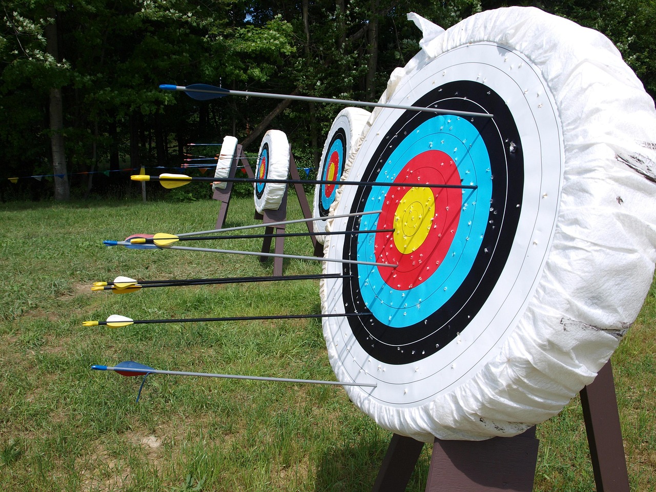
At the 2026 State Games of America, organizers merged two archery tournaments into a single 10 hour day. Anne Abernathy, 73, was the only archer who attempted both. She credits her gold metal in archery to photobiomodulation.
She won both, and posted the highest recurve score of the day of any archer on the field, male or female. Archery asks for exactly what people are told to expect less of with age: sustained attention, precise repetition, composure under pressure.
Anne shot 204 arrows across 10 hours in the heat, and put five of her final six in the gold. Her score rose to 747, up from 735 at her last competition in 2025
The Vielight Neuro has been part of Anne’s daily routine through a year of rehabilitation and her return to competition.
“I would not have attempted a challenge of this magnitude without Vielight.”
What in the world are secrets to living a long, healthy and happy life? Ninety year old Reg MacDaniel MD offers his answer to this most important question. His research has revealed that a natural extract from the Aloe plant called Acemannan provides remarkable healing for a variety of chronic conditions.
What are the best remedies for chronic conditions? Many centuries ago Hippocrates knew the answer to this question:
“Let your food by your medicine and your medicine be your food.”
Food is the best medicine when compared to all the options. It is a natural remedy with no side effects that is more effective than taking dozens of supplements every day.
Of course it depends on what food we are talking about here. The typical American diet acidifies the body which leads to chronic illnesses like Parkinsons. The ability to buffer acidity is exhausted by the young age of 45.
Worse, acidic bodies significantly reduce oxygen levels. Without sufficient oxygen circulating through the body, chronic illness can not be reversed.
Dr. Reg has played a central role in identifying and extracting a substance from the aloe vera plant (Acemannan) that boosts the immune system and reduces inflammation. Both issues are well known to be a cause of chronic conditions. Acemannan is a star player in cellular synthesis. It forms a membrane that promotes cell harmony and balance, a state known as homeostasis.
Dr. REg devotes all of his time and energy to spreading the word about the health benefits of Acemannan and organizing studies to evaluate its effects.
There is no doubt about it. The most effective medicines with no side effects are derived from plants. Extracts from certain foods heal a variety of conditions including Parkinsons and Alzheimers. They do not address neurological symptoms specifically but provide support for the body’s ability to heal from the inside out rather than suppressing symptoms temporarily.
We all know on some level that the aloe plant heals burns and cuts, but the health benefits from a specific extract derived from Aloe turn out to be far greater than wound healing alone.
Researchers have named this extract Acemannan. Its health benefits are considerable including it …
Reduces inflammation
Has a molecular structure similar to a mother’s milk which offers support needed by newborns to thrive
Helps regulate the immune system by stimulating one that is under-active and depressing one that is over-active
Supports the function of a healthy gut
Repairs cellular damage
Facilitates the creation of new stem cells
Stimulates the production of glutathione
As a natural food the extract of Acemannan from the Aloe plant has no documented side effects.
Visit the replay link below of a presentation by Kari Skinner who discusses the healing properties of Acemannan. Her focus is on how this extract in particular offers the potential to help persons currently confronting neurological symptoms.
I recommend this natural treatment option for Parkinsons be given serious consideration. It supports the ability of the body to heal rather than simply suppressing symptoms with medicines and supplements for the rest of a person’s life.
Pharmaceutics. 2023 Jul 9;15(7):1913. A New Biomaterial Derived from Aloe vera—Acemannan from Basic Studies to Clinical Application
Abstract
Aloe vera is a kind of herb rich in polysaccharides. Acemannan (AC) is considered to be a natural polysaccharide with good biodegradability and biocompatibility extracted from Aloe vera and has a wide range of applications in the biomedical field due to excellent immunomodulatory, antiviral, antitumor, and tissue regeneration effects.
In recent years, clinical case reports on the application of AC as a novel biomedical material in tissue regenerative medicine have emerged; it is mainly used in bone tissue engineering, pulp–dentin complex regeneration engineering, and soft tissue repair, among other operations. In addition, multiple studies have proved that the new composite products formed by the combination of AC and other compounds have excellent biological and physical properties.
Opportunity expires today. Consultations with Founder of Parkinsons Recovery Robert Rodgers PhD to help you sort out the natural options that would prove most helpful to your Recovery.
* Is your recovery stuck in the mud? * Are you totally frustrated with symptoms that are getting worse?
Turn your recovery around with ongoing one on one consultations with me to identify the best treatment options that will address your situation. Visit the link below for more information and to register.
Traditional mouthwashes are thin liquids, filled with alcohol and harsh chemicals that burn the mouth. Alcohol destroys the good bacteria needed to digest food. For these reasons a natural remedy for gut problems is not mouthwashes that contain alcohol.
SUVIA is entirely different. SUVIA isn’t just another mouthwash. It is a daily oral care RINSE designed to support a healthier mouth.
It all comes down to the hydrogel difference. A hydrogel is a three-dimensional network of polymer chains that retain massive amounts of water—often up to 99% of their weight—while maintaining a solid or gel-like structure
SUVIAforms a protective shield. Swishing it creates an ultra thin, hydrogel film. You won’t see it, but the film stays in place, adhering to oral tissue to manage any discomfort. It also protects wounds.
SUVIA is made of 100% natural food ingredients made from aloe vera, xylitol, cinnamon, clove, and thyme and water. No alcohol. No synthetic chemicals.
SUVIA creates the conditions for optimal healing. Unlike mouthwashes, it does not numb the mouth. Instead, it manages pain while maintaining the moist environment required for optimal wound healing.
SUVIA manages pain/discomfort and protects oral wounds from further irritation and contamination to promote optimal healing from a variety of conditions including …
Dentures
Braces & Appliances
Cold Sores
Fever Blisters
Canker Sores
Implants
Brushing and Flossing
Post Dental Work
Eating and Drinking Injuries from hot or sharp food
Any other issued that cause pain, discomfort or blood in your mouth
Conclusion
A reason people have gut problems and constipation that defy resolution centers on problems that reside in the mouth, not the digestive system itself. Heal problems in your mouth to celebrate a natural remedy for gut problems. And when the gut is back on line, neurological symptoms should gently resolve.
New technologies offer exciting opportunities for persons confronting chronic conditions like Parkinsons. Six ground breaking discoveries that heal chronic conditions are revolutionizing treatment options for persons experiencing neurological problems like Parkinsons and Alzheimers.
Available for a limited time only. Consultations with Robert to Facilitate Your Recovery
* Is your recovery stuck in the mud? * Are you totally frustrated with symptoms that are getting worse?
Turn your recovery around with a one on one consultation with me to identify the best treatment options that will address your situation. Visit the link below for more information and to register.
Good evening Robert. I’ve been reading these great books written by a guy named, Robert Rodgers, PhD…have you heard of him? I want everyone to know about how energy healing calms tremors or at least that has been my experience.
I was reading about energy healing, amongst a myriad of protocols and treatments suggested in your top ranked book Road to Recovery from Parkinsons Disease. I proceeded to type in a google search: energy healing in Hollywood for free.
Long story short…there is a spiritual community that is literally 5 minutes from where I live, and every Tuesday night, they welcome people for 1/2 hr hands on energy healing…cost: zero.
They train these healers and it’s all very legit. So I called, made an appointment and Tuesday night I went. Sat erect in a chair with my hands on my legs. My right hand tremor was very apparent. 10 minutes in, my fingers stopped shaking…my thumb continued. Then, at 15 minutes…my entire hand stopped shaking for the duration of the session, and it didn’t come back till I was outside. Amazing! Energy healing calms tremors.
The best news is that they suggested me coming every Tuesday night indefinitely!! Robert, it’s true…
I am guessing you have likely never heard about redox signaling molecules. A search on the depository of medical publications (pubmed.gov) reveals that more than 400 studies have been published in the first six months of 2026 alone. Over 7,300 studies have been published overall. This is a recent discovery that has attracted the interest of hundreds of researchers across the globe. I want you to know more about it too!
As is evident from my presentation above, I am excited about the potential for redox signaling molecules to offer welcome relief from neurological challenges.
Get all of your questions answered in several ways. First, visit https://www.recoverwithredox.net. Complete the contact form and a representative from the company will contact you to answer all of your questions.
Second watch this marvelous presentation on redox signaling by Alan Noble who really is the first person who recognized the value of this technology to bring the body back into balance and harmony.
Third, watch this short 6 minute youtube video that demonstrates how redox signaling has a quick impact on reducing inflammation in the brain.
What success have users had from using redox signaling? A website offers interviews with individuals who report positive experiences. Visit: realredoxresults.com. Enter the password redox for access to the interviews.
In summary, I recommend you take this option under serious consideration. It has clearly helped many people and offers the opportunity to help you too.
This brief post explores the connection between trauma and Parkinson’s disease. An idea that has been floating around for decades is that we are stuck with the genes we are born with. Some people are lucky and some unlucky. If we have a gene that predicts cancer – it is likely we will get cancer. If we have the genes that are disposed to Parkinson’s disease, we are likely to develop the symptoms of Parkinson’s. Some of us are dealt a lousy hand in life.
What circumstance switches a gene on or off? The answer will fascinate you. It is trauma! Research shows that the tendency toward post traumatic stress (which exhibits symptoms similar to those of Parkinsons) are passed down to the next generation. Children of holocaust survivors pass down a predisposition of stress and anxiety to their offspring.
You may have always thought that there have been no significant traumatic events in your life that could account for the degree of stress and anxiety that you currently experience. When you have heard me talk about the critical role of stress and trauma in affecting Parkinson’s symptoms, you may well have dismissed my argument. Recent research clearly shows that the origins of stress can reside in the experiences of ancestors.
How could traumatic events influence whether genes are turned on or off? Simple. When something cataclysmic happens to people they will say that they are not the same person. You may well have heard yourself or friends say the same thing. There it is my friend.
Robert Rodgers PhD
Founder 2004
Parkinsons Recovery®
https://www.parkinsonsrecovery.com
robert@parkinsonsrecovery.com
Olympia Washington
877-526-4646
This post explores why high dose thiamine therapy can offer symptom relief from some persons diagnosed with Parkinsons.The good news of the day is that there may well be a simple solution to debilitating symptoms of Parkinsons for some individuals.
Some medical professionals endorse high dose thiamine therapy as a treatment for Parkinsons. Studies have reported that Thiamine (which is the technical term for Vitamin B1) tends to be deficient in persons experiencing neurological symptoms. The deficiency in part is due drug depletions from certain medications used to treat Parkinsons.
Beriberi is a disease of the nervous system which is also due to a deficiency of Thiamine (Vitamin B1). Thiamine is needed to breakdown food into glucose. It is also found in neural tissues.
Some people with Parkinsons celebrate a welcome improvement in symptoms after getting high dose B1 therapy from their doctor. Others have not.
The original source that recommended high dose B1 therapy is Dr. Anthony Costontini. You can listen to my interview with him here where he details his reasons for recommending this simple treatment.
How can you know whether high dose B1 therapy might be helpful for you? A good clue is whether you experience the symptoms associated with Beriberi disease which are similar to some of the symptoms of Parkinsons.
Beriberi Symptoms of the Nervous System
Tingling or loss of feeling in the hands and feet
Muscle weakness and pain in the legs
Difficulty walking or loss of balance and coordination
Lower leg paralysis or foot drop
Do you experience any of the symptoms above? If so, high dose thiamine therapy might just provide the relief you seek. I suspect this is why some people celebrate such welcome relief because they are deficient in vitamin B1. Consider discussing this treatment option with your doctor.
Research on B1 Deficiencies and Beriberi Disease
Curr Nutr Rep. 2026 Jan 29;15(1):8. B Vitamin Deficiencies and Associated Neuropathies
Abstract
Purpose of review: This review examines both historical and recent evidence to clarify the current understanding of the relationship between B vitamin deficiencies and neuropathies.
Recent findings: Vitamin B1 (thiamine) deficiency can lead to neurological disorders such as beriberi and Wernicke’s encephalopathy, conditions with significant prevalence and mortality rates. Vitamin B12 (Cobalamin) is crucial for DNA synthesis, fatty acid metabolism, and myelin production, with its deficiency leading to neuropathies and cognitive disorders. Excess vitamin B6 (pyridoxine), rather than deficiency, appears to be associated with neuropathy.
Takeaways/conclusions: Vitamin B1 and B12 deficiencies are linked to classic neuropathies, while the connection between vitamin B6 deficiency and neuropathy is less clear, though excess B6 is associated with neurotoxicity. Nutritional deficiencies are less common in developed countries but remain significant in developing nations. In developed countries, factors like alcohol consumption, bariatric surgery, and metformin use are increasing these deficiencies in clinical practice.
******
Med Princ Pract. 2026;35(1):18-24. An Overview of Beriberi
Abstract
Beriberi is a nutritional disorder caused by thiamine deficiency. Classically, Beriberi presents in two primary clinical forms: “wet” Beriberi, which features heart and circulatory system impairment, and “dry” Beriberi, which causes polyneuropathy.
Polyneuropathy is damage to peripheral nerves which are located outside your brain and spinal cord. The condition usually starts in the feet and hand and causes numbness, tingling, and weakness.
Although it is an easily treatable condition, it is often misdiagnosed and can be life-threatening if not promptly recognized and managed. The diagnosis of Beriberi is performed by the signs and symptoms of the disease and can be confirmed by thiamine deficiency identification or by therapeutic testing.
Immediate thiamine administration in the presence of clinical manifestations of the disease is recommended. Overall, 100-300 mg daily doses are enough to improve symptoms.
The big picture when it comes to understanding of the connection between sleep and Parkinsons is a gross imbalance between the sympathetic nervous system and the parasympathetic nervous system.
Flight or Fight?
The sympathetic system is easily understood as a flight-fight response. When we are afraid or anxious, we want to either run away from the problem or fight it directly.
Rest and Digest
The parasympathetic system is the opposite – the rest and chill out digest system. No one can digest their food when they are running a marathon. And no one can get to sleep if their muscles are postured to fight off an unwanted intruder into the bedroom.
We survive because both systems serve vital functions. When neurological symptoms become problematic, the sympathetic fight-flight system takes charge most of the time. Adrenaline and cortisol are fueling the fire inside out tissues. We are forever ready to engage and fight, seldom chilling out to rest and digest.
When you calm down your stress level and release past traumas you awaken a dormant parasympathetic nervous system. We all can get overly excited in the flash of an instant. Relaxing can take endless hours of meditation, soft music and pleasant company.
Connection Between Sleep and Parkinsons is Profound
The formidable challenge confronted by anyone experiencing the symptoms of Parkinsons confront is that rarely if do they drop down in the deep REM sleep that is critical for healing. Healing is successful only when we experience the level of deep sleep that is critical for rejuvenating and regenerating tissues.
A persistent flight flight sympathetic nervous system that dominates during the day does not relinquish control at night when sleeping. Nope. Once the sympathetic nervous system shifts into control is is not about to give it up. The refusal to back down is analogous to politicians who refuse to relinquish their power.
We slip down into that unconscious state of deep sleep when the body is literally paralyzed (in a good sense). There is a switch in the mid brain which is designed to turn on and off this ability to become paralyzed.
With Parkinson’s, the on-off switch gets stuck.
Many people with Parkinsons symptoms hold the belief they are getting good enough sleep. Most people with neurological challenges are not. are not.
The bottom line: introduce into your recovery program a set of therapies and ways that flip your switch at night so that your sympathetic nervous system is deactivated. Recovery will be at a stalemate until you achieve the deep state of REM sleep night after night.
What makes a difference between people who recover from Parkinson’s disease and those who do not? Eating a healthy, clean diet plays a leading role. Processed food destroys gut health.
People who eat processed foods with toxic additives tend flop with their recovery
People who eat live, organic unprocessed diets thrive with their recovery.
Diet is key.
In my over 1,000 discussions with persons diagnosed with Parkinson’s disease, I have encountered many who are well aware of this truth and make valiant efforts to clean up their diets, but with only partial success. Why is this?
They are addicted to those diet colas and ice creams and other processed foods. The following research will offer a compelling reason why the addictions have to be overcome for recovery to manifest.
Processed Food Destroys Gut Health
There are “good” and “bad” bacteria in the gut. The many different strains of good bacteria are necessary for healthy digestion. The bad bacteria and more virulent strains can gobble up the good bacteria.
Many people eat relatively clean diets but little nutrition is absorbed because the bad bacteria are in charge of their digestion. Good bacteria is lacking and even absent in some. Unfortunately the result is constipation and a variety of other digestive challenges for many persons who experience Parkinson’s symptoms.
Recent evidence indicates that the “bad” bacteria are beginning to dominate the “good” in research samples that have been studied. Why is this?
Researcher Robert Britton PhD from the Baylor College of Medicine and his colleagues wanted to know why the more virulent strains like clostridium difficile or C.diff (which is horrible, life threatening infection) were becoming so much more prevalent and knocking out the good bacteria.
These horribly nasty bacteria love to eat some of the nastiest “bad” additives used to extend the shelf life and moisture of processed foods like diet colas. One of the worse culprits is the sugar trehalose which the bad bacteria loves to eat.
The bottom line:
Continue to eat any processed foods and diet colas and your digestion will implode. Constipation will persist. Long term prognosis is the worst imaginable.
When and if you fall off the wagon and drink that one little diet cola or eat that “delicious” processed brownie you will pay a dear price. Recovery ain’t happen’ for you my friend.
I do understand why it is difficult to break addictions to eating processed foods. There is always an underlying emotional attachment to them. But, if you have set the intention to reverse whatever symptoms you currently experience that are associated with a diagnosis of Parkinson’s disease, you do not have a choice.You have to eat a 100% clean, organic, unprocessed food.
The answer to the question “What brand of CoQ10 is best?” has puzzled me for many years. The challenge with finding a reliable source turns on the fact Coq10 is not absorbed by the body.
I have been telling members of my global audience to avoid purchasing inexpensive brands because you are throwing your money away. The more expensive brands are clearly preferable.
The melting point of CoQ10 is 10 degrees above normal body temperature. Most of the supplements you buy – even those in oil – have crystallized even before you put them in your body.
Check it out for yourself. Paste whatever brand of CoQ10 you currently take on a sheet of wax paper. Then, shine the light from flashlight (or your phone) onto the spread of CoQ10. Do you see crystals? If so, your body is likely absorbing a tiny fraction of the CoQ10 from the supplement you are taking – as little as 1%.
In short, you are throwing your money down the toilet. This is partly why research studies find that you need to take relatively large doses of CoQ10. If the absorption is so terribly low, a high dose would be necessary.
You are having to spend a lot of money to get a modest benefit in return.
Natural pharmacist Ross Pelton tells me that the story does not end there. Even if the CoQ10 has not crystallized before you take it, it may crystallize after you take it. It was Ross who introduced me to the 75 studies of the Pharmanord brand of CoQ10 which reports a very high absorption rate.
I called the company to explore this further. I acquired some for myself and my family and have experienced more energy which I need desperately to keep up with the demands of Parkinsons Recovery. After over a decade of hesitation to recommend a best CoQ10 brand, I am finally comfortable recommending this one.
The price is also reasonable and you do not have to take as much. You can order the PharmaNord (https://www.pharmanord.com) CoQ10 directly on their website. Their professional staff set up a 20% discount for members of my audience. Enter the coupon code PRQ10WEB to claim a 20% discount (not case sensitive) if you are shipping to a USA destination.
Check out the Pharmanord research for yourself. It is extensive and impressive.
Caloric restriction is a lifestyle modification in which the amount of calories ingested is reduced to a significant amount without resulting in malnutrition. The studies posted below conclude caloric restriction is beneficial for Parkinsons.
Caloric Restriction Mimetics offer a way to get the benefits of calorie restriction without the difficulty of strictly limiting food intake. These compounds act in ways similar to actual calorie reduction by activating cellular cleanup without requiring severe dietary limits.
The compounds are similar to actual calorie reduction by activating cellular cleanup without requiring severe dietary limits.
Key examples include spermidine (found in mushrooms) and resveratrol (found in grapes and berries). They mimic fasting, boost cell repair and offer potential treatments for neurodegenerative conditions like Parkinsons. They also improve glucose metabolism found to be an issue associated with symptoms of Parkinsons.
Summary: Caloric Restriction is Beneficial Parkinsons and show compelling promise as a natural therapy for all neurodegenerative conditions.
Why Caloric Restriction Matters
Neuroscience. 2025 Feb 16:567:235-248. Mechanisms and early efficacy data of caloric restriction and caloric restriction mimetics in neurodegenerative disease
Abstract
Neurodegenerative disorders (NDDs) have been prevalent for more than a decade, and the number of individuals affected per year has increased exponentially. Among these NDDs, Alzheimer’s disease, which causes extreme cognitive impairment, and Parkinson’s disease, characterized by impairments in motor activity, are the most prevalent.
While few treatments are available for clinical practice, they have minimal effects on reversing the neurodegeneration associated with these debilitating diseases. Lifestyle modifications and dietary choices are emerging and promising approaches to combat these disorders.
Of the lifestyle changes that one could adopt, a major habit is caloric restriction. Caloric restriction is a lifestyle modification in which the amount of calories ingested is reduced to a significant amount without resulting in malnutrition. However, maintaining such a lifestyle is challenging.
As alternatives, certain compounds have been recognized to mimic the effects produced by caloric restrictions. These compounds are called caloric restriction mimetics (CRMs). Among these compounds, some have been designated established CRMs, namely, resveratrol, metformin, and rapamycin, whereas several other candidates are termed potential CRMs because of a lack of conclusive evidence of their effects.
The potential CRMs discussed in this review are quercetin, chrysin, astragalin, apigenin, curcumin, epigallocatechin-3-gallate, and NAD+ precursors. This review aims to provide an overview of these CRMs’ effectiveness in preventing neurodegenerative disorders associated with aging. Moreover, we highlight the clinical relevance of these compounds by discussing in detail the results of clinical trials on them.
******
Curr Nutr Rep. 2024 Dec;13(4):691-700. Effects of Caloric Restriction and Intermittent Fasting and Their Combined Exercise on Cognitive Functioning: A Review
Abstract
Purpose of review: The impact of dietary habits on cognitive function is increasingly gaining attention. The review is to discuss how caloric restriction (CR) and intermittent fasting (IF) can enhance cognitive function in healthy states through multiple pathways that interact with one another. Secondly, to explore the effects of CR and IF on cognitive function in conditions of neurodegenerative diseases, obesity diabetes and aging, as well as potential synergistic effects in combination with exercise to prevent cognitively related neurodegenerative diseases.
Recent findings: With age, the human brain ages and develops corresponding neurodegenerative diseases such as Alzheimer’s disease, Parkinson’s disease, and epilepsy, which in turn trigger cognitive impairment. Recent research indicates that the impact of diet and exercise on cognitive function is increasingly gaining attention.
The benefits of exercise for cognitive function and brain plasticity are numerous, and future research can examine the efficacy of particular dietary regimens during physical activity when combined with diet which can prevent cognitive decline.